Hip Muscles Diagram - Hip Pain Explained Including Structures Anatomy Of The Hip And Pelvis. Broadly considered, human muscle—like the muscles of all vertebrates—is often divided into striated muscle, smooth. The muscular system is responsible for the movement of the human body. *click them to make them larger & view details. Related online courses on physioplus. See more ideas about muscle diagram, medical anatomy, body anatomy.
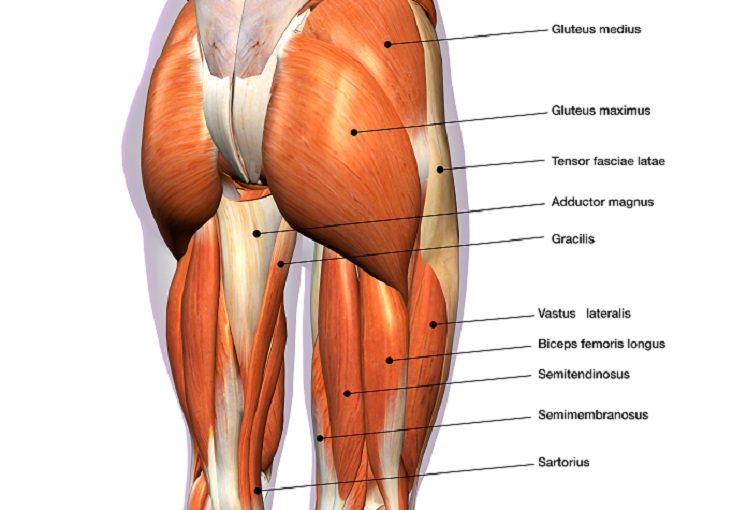
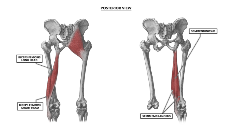
Broadly considered, human muscle—like the muscles of all vertebrates—is often divided into striated muscle, smooth. There are anterior muscles diagrams and posterior muscles diagrams. Smartdraw includes 1000s of professional healthcare and anatomy chart templates that you can modify and make your own. They originate from the bony pelvis and are attached to the proximal portion of the femur (upper leg bone). Gluteus maximus, biceps femoris, semitendinosus, semimembranosus at the back and the.
Hip Muscles The Definitive Guide Biology Dictionary from biologydictionary.net These two muscles produce lateral rotation at the hip and are innervated by the obturator internus and quadratus femoris nerves. There are anterior muscles diagrams and posterior muscles diagrams. Start studying book interior hip muscles. The main muscles of the hip and pelvis consistsof the iliopsoas, pectinues, rectus femoris and sartorius at the front. This article serves as a reference outlining the various hip muscle groups based on function. Gluteus maximus, biceps femoris, semitendinosus, semimembranosus at the back and the. The pelvic floor muscles provide foundational support for the intestines and bladder. These muscles are separate in the abdomen, but they join.
Now that you watched the video, you.
Review muscle diagram using the 2 diagrams below: Knee assessment and hip mechanics online course: This diagram depicts hip muscles diagram and explains the details of hip muscles diagram. Almost every movement in the body is the outcome of muscle contraction. *click them to make them larger & view details. The hip and pelvic muscles include: The following diagram illustrates the actions of the terms adduction, abduction, flexion and extension at the different joints. Knee assessment and hip mechanics learn how hip and pelvis. 25.09.2020 · the hip muscles encompass many muscles of the hip and thigh whose main function is to act on the thigh at the hip joint and stabilize the pelvis.without them, walking would be impossible. Human muscle system, the muscles of the human body that work the skeletal system, that are under voluntary control, and that are concerned with movement, posture, and balance. The hips also enable people to lift their feet two individual muscles called the psoas major and the iliacus form the iliopsoas muscle. Smartdraw includes 1000s of professional healthcare and anatomy chart templates that you can modify and make your own. Related online courses on physioplus.
Review muscle diagram using the 2 diagrams below: Now that you watched the video, you. The sacrum bone is almost always noticeable, no matter what the body type, because it is not covered with muscles or substantial fatty tissue. Start studying book interior hip muscles. Their main function is contractibility.
Crossfit Hip Musculature Part 2 Posterior Muscles from www.crossfit.com Smartdraw includes 1000s of professional healthcare and anatomy chart templates that you can modify and make your own. They can be divided into three main groups: The following diagram illustrates the actions of the terms adduction, abduction, flexion and extension at the different joints. The gluteus medius, gluteus minimus, piriformis, tensor fasciae latae on the outside. Flexors, extensors, adductors, abductors, lateral rotators. 25.09.2020 · the hip muscles encompass many muscles of the hip and thigh whose main function is to act on the thigh at the hip joint and stabilize the pelvis.without them, walking would be impossible. The muscular system is responsible for the movement of the human body. Human muscle system, the muscles of the human body that work the skeletal system, that are under voluntary control, and that are concerned with movement, posture, and balance.
These muscles are separate in the abdomen, but they join.
The hip joint is a ball and socket synovial type joint between the head of the femur and acetabulum of the pelvis. This diagram depicts hip muscles diagram and explains the details of hip muscles diagram. The hips are the central pivot point of the entire body, supporting its weight during movement and when standing. Depending on the situation, with the pelvis in a fixed position the muscles move around the thigh. Diagram representing the anterior view of the muscle groups adductor brevis, adductor longus and adductor magnus. The major muscles that produce movements of the hip joint are categorized into functional groups; It therefore serves the artist as a dependable visual landmark for the location of muscular forms. The sacrum bone is almost always noticeable, no matter what the body type, because it is not covered with muscles or substantial fatty tissue. See more ideas about muscle diagram, medical anatomy, body anatomy. Flexors, extensors, adductors, abductors, lateral rotators. Start studying book interior hip muscles. Their main function is contractibility. This is important to understand the actions of the thigh muscles in limb anterior compartment thigh muscles.
The hips also enable people to lift their feet two individual muscles called the psoas major and the iliacus form the iliopsoas muscle. Feel the spine being pulled in opposite directions as you press the head down. The sacrum bone is almost always noticeable, no matter what the body type, because it is not covered with muscles or substantial fatty tissue. Diagram representing the anterior view of the muscle groups adductor brevis, adductor longus and adductor magnus. The gluteus medius, gluteus minimus, piriformis, tensor fasciae latae on the outside.
Groin Muscles Diagram Koibana Info Leg Muscles Diagram Hip Muscles Anatomy Leg Muscles Anatomy from i.pinimg.com Knee assessment and hip mechanics learn how hip and pelvis. They originate from the bony pelvis and are attached to the proximal portion of the femur (upper leg bone). Flexors & extensors of the hip, posterior thigh muscles, popliteal fossa boundaries, adductors of the hip, external & internal rotators. Flexors, extensors, adductors, abductors, lateral rotators. Smartdraw includes 1000s of professional healthcare and anatomy chart templates that you can modify and make your own. This is important to understand the actions of the thigh muscles in limb anterior compartment thigh muscles. See more ideas about muscle diagram, medical anatomy, body anatomy. Diagram representing the anterior view of the muscle groups adductor brevis, adductor longus and adductor magnus.
Their main function is contractibility.
Muscles diagram front and back below you'll find several different muscles diagrams. Knee assessment and hip mechanics learn how hip and pelvis. An important group of muscles in the pelvis is the pelvic floor. Depending on the situation, with the pelvis in a fixed position the muscles move around the thigh. This diagram depicts hip muscles diagram and explains the details of hip muscles diagram. Other muscles also assist in the abduction of the thigh at the hip joint, but they do not belong to the abductor group. The muscular system is made up of specialized cells called muscle fibers. Label the major muscles of the body. Gluteus maximus, biceps femoris, semitendinosus, semimembranosus at the back and the. Their main function is contractibility. Diagram representing the anterior view of the muscle groups adductor brevis, adductor longus and adductor magnus. Muscles, connected to bones or internal organs and blood vessels, are in charge for movement. Attached to the bones of the skeletal system are about 700 named.
Share :
Post a Comment
for "Hip Muscles Diagram - Hip Pain Explained Including Structures Anatomy Of The Hip And Pelvis"


Post a Comment for "Hip Muscles Diagram - Hip Pain Explained Including Structures Anatomy Of The Hip And Pelvis"